
Cody's Headshaking Case Study
A whole-horse approach to a complex presentation
Cody is a 16-year-old Quarter Horse with a three-year history of seasonal headshaking. His signs were normally at their worst during spring and autumn and included sudden head flicking, twitching, nose rubbing and clamping of the left nostril when triggered. Wind was a particularly clear aggravating factor.
By the time I first assessed Cody on 8 August 2026, his owners had already pursued extensive veterinary investigation. This included sinus scoping, a full diagnostic work-up, previous gastric investigation and a bone scan. The bone scan had identified increased activity around a sacroiliac joint and a hock, with low-grade arthritic change. He was receiving ongoing veterinary management, and acupuncture had previously helped reduce twitching around his withers.
The remarkable part of Cody's progression was not simply that he improved. It was that he entered September and October, normally his most difficult period, with virtually no headshaking signs.
A history of trauma and compensation
Cody also had a significant trauma history. At four years old, he pulled a fence post while attached by his headcollar. Approximately six months before my first assessment, he fell from a trailer while still attached.
His owners felt that the head and neck trauma might be relevant. However, with a complex case like this, it is rarely helpful to search for one structure to blame. My role was to assess how Cody's cranium, jaw, neck, thorax, spine, pelvis, limbs and visceral relationships were functioning together, while veterinary investigation continued to exclude or manage pathology.
What I found at the first session
Cody presented with extensive layered compensation.
His upper neck and cranial system were markedly restricted. The first cervical vertebra, C1, was significantly rotated, and the relationship between the hyoid, mandible, temporomandibular joints and temporal bones was heavily decompensated on both sides. The cranial base retained a complex strain pattern, with bilateral compression around the spheno-petrosal and occipitomastoid relationships. Sensory testing also suggested altered responsiveness through the upper left trigeminal field, alongside congestion through the upper nasal region.
The restriction was not confined to his head. He showed:
- extensive restriction through T18 and the lumbar spine
- a collapsed sternum and weak thoracic sling
- marked bracing through the thoracic outlet and thoracolumbar junction
- restricted ribs and an asymmetrical first-rib pattern
- a right radial carpal bone restricted in flexion
- a complex sacral torsion with compensatory and decompensatory patterns through both ilia, hips, stifles and hocks
- tension through the left kidney, gelding scar, prostate, right spermatic cord and internal inguinal ring
Rather than viewing each finding in isolation, I followed the relationships between them. The right pelvic complex, left hip and bilateral jaw-temporal relationships appeared to have become important compensatory points along the fascial chains. Restricted thoracic expansion and breathing mechanics were likely adding further strain through the lumbar region.
The first treatment
Treatment was adapted to Cody's tolerance and included structural, fascial, craniosacral and visceral approaches. I worked through the restrictions found during assessment, including the cranial base, temporals, TMJs, upper cervical spine, thoracic outlet, sternum, ribs, thoracolumbar region, pelvis and relevant visceral relationships.
Cranial treatment included a tentorium lift and drainage work. Cody was then given two days off before gradually returning to appropriate work, with his owners monitoring his symptoms closely.
Six weeks later
Cody was reassessed on 23 September 2026. His owners described the improvement as amazing.
September and October were normally his worst months, yet they had seen:
- no nose rubbing
- no head flicking
- no facial twitching
- far less tension through the upper neck
- improved ability to perform groundwork and stretch his neck
They had needed his ceramic ear mask only once, on a particularly windy day. Most notably, the signs stopped immediately when the mask was fitted. Previously, once an episode had begun, putting the mask on would not stop it.
This was a meaningful functional change, although it is important to place it in context. Cody had also received veterinary treatment to his hocks and sacroiliac region approximately four weeks before the reassessment. His progress therefore belongs within a collaborative picture rather than being attributed to one intervention alone.
The second assessment
The second-session findings were almost completely different from those of the first appointment. For me, this was encouraging: the original pattern had shifted rather than simply returning unchanged.
There was still some cranial tension, but substantially less. The principal remaining pattern centred on C1, the left temporal and TMJ, and the maxillary or V2 division of the trigeminal nerve. Releasing C1 produced an immediate release through the left C4-C5 region.
The left scapula remained in a downslip position and was accepting considerable weight. T2-T3 and the surrounding tissues were significantly restricted. The lumbar emphasis had moved from T18-L2 to L3-L4, and I addressed associated bladder and prostate relationships before adjusting the local spinal restrictions.
The hindquarter was much improved overall. Reduced inflammation following veterinary treatment allowed me to assess and treat the hocks and cuboids more effectively. The sacroiliac region remained mechanically compromised, with a left-axis, left-rotation sacral pattern and decompensation through both ilia, but it responded very well to treatment.
Cody was exceptionally receptive throughout. By the end of the session, the superficial fascial contours through his neck, shoulder, ribcage, thoracolumbar region and pelvis looked visibly smoother, almost as though they had been ironed out.
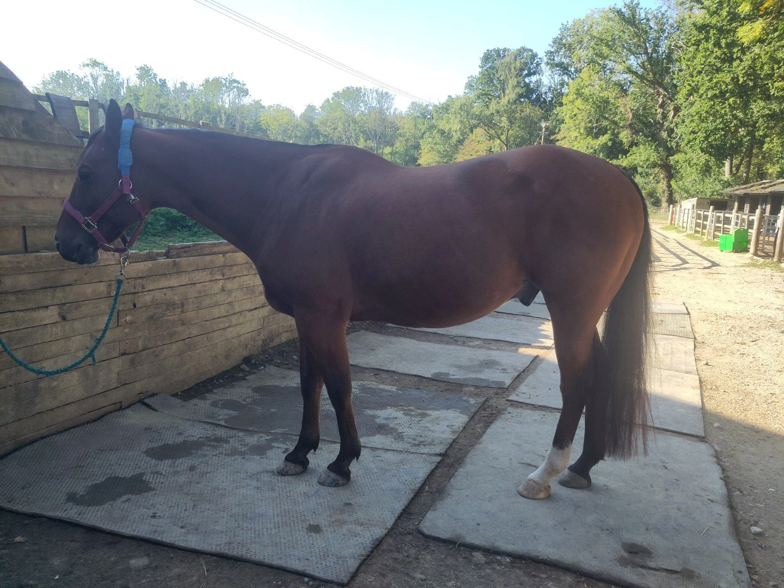
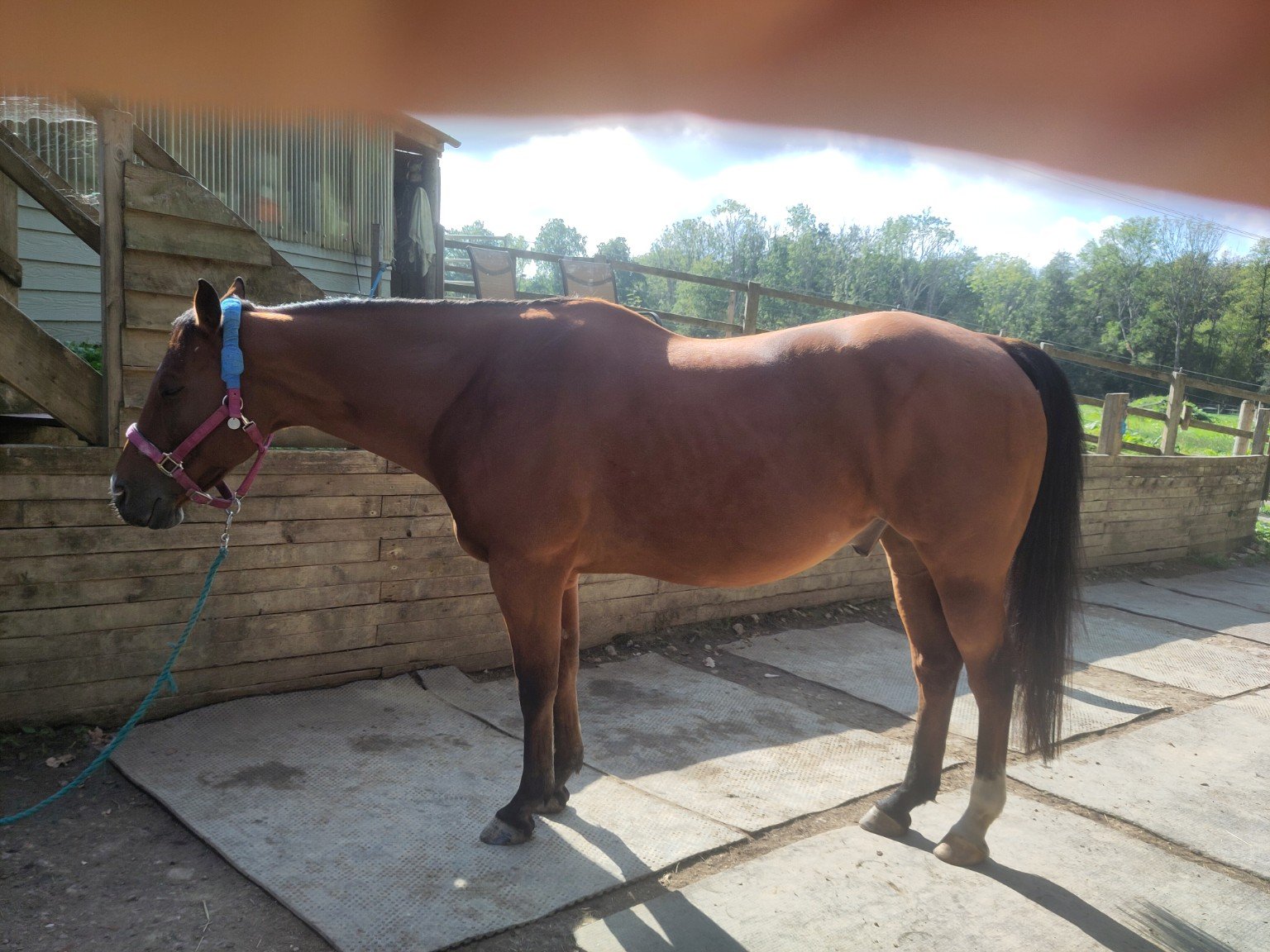
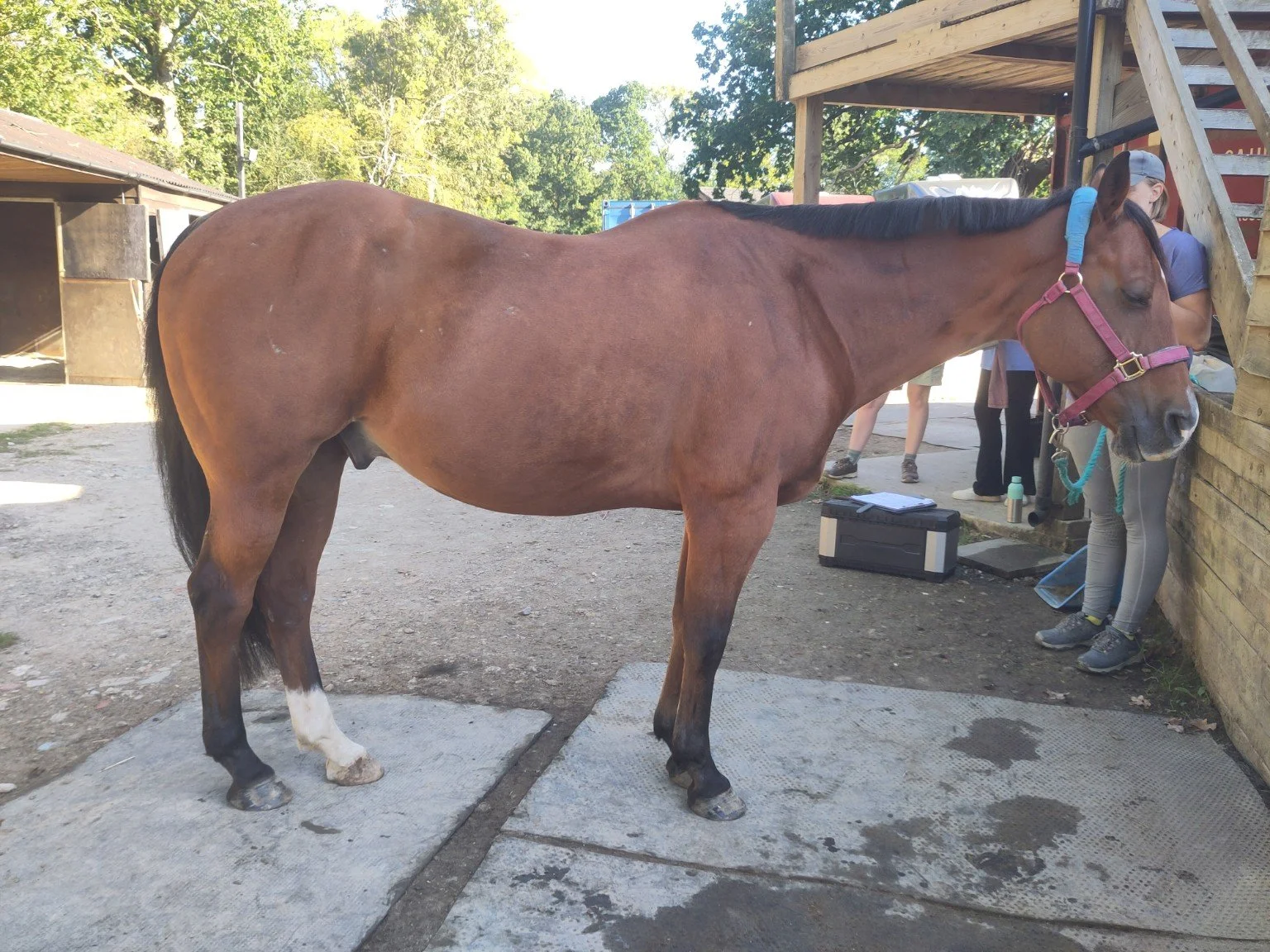
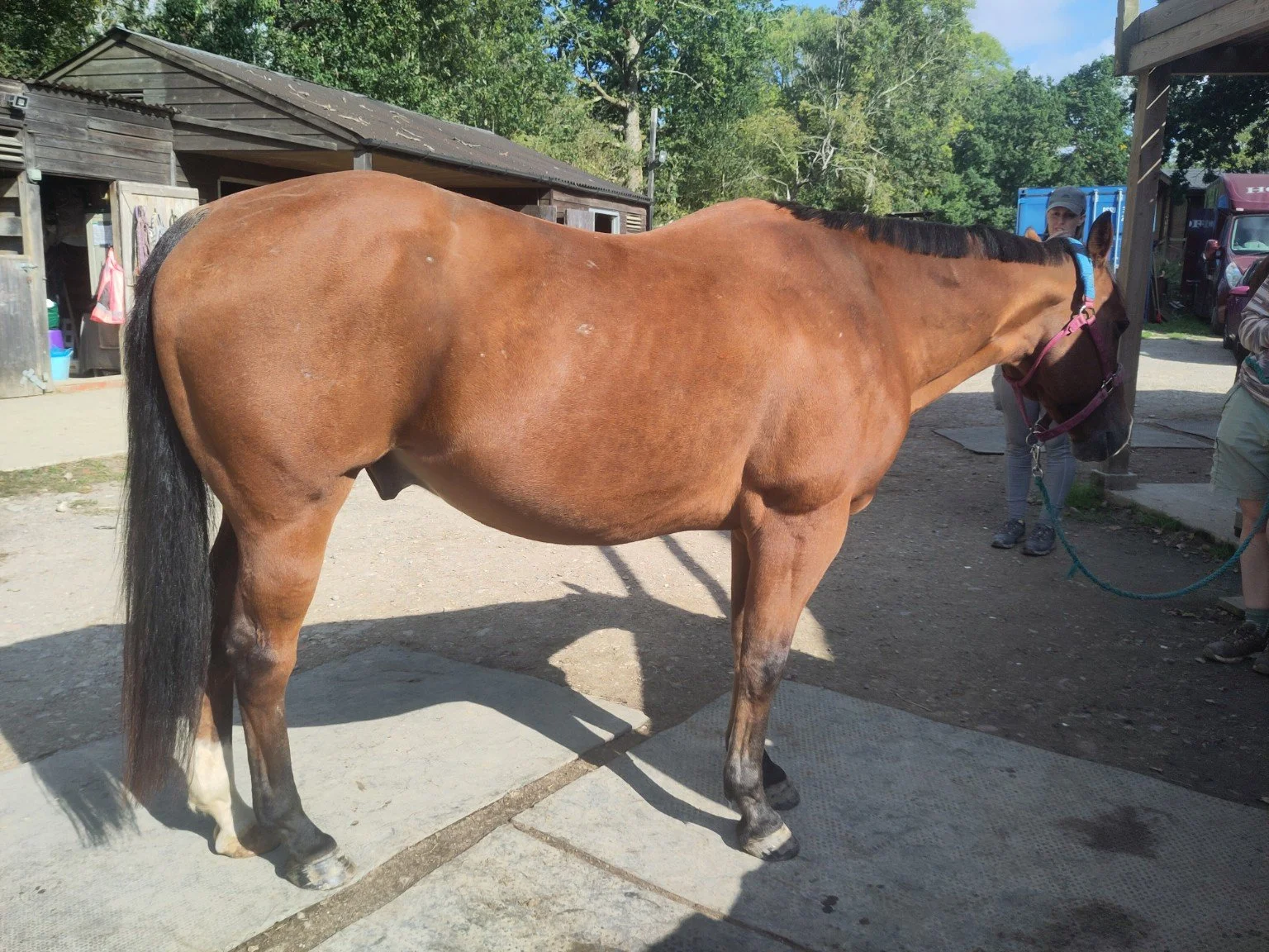
Before and after photographs
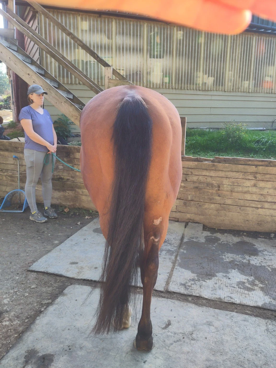
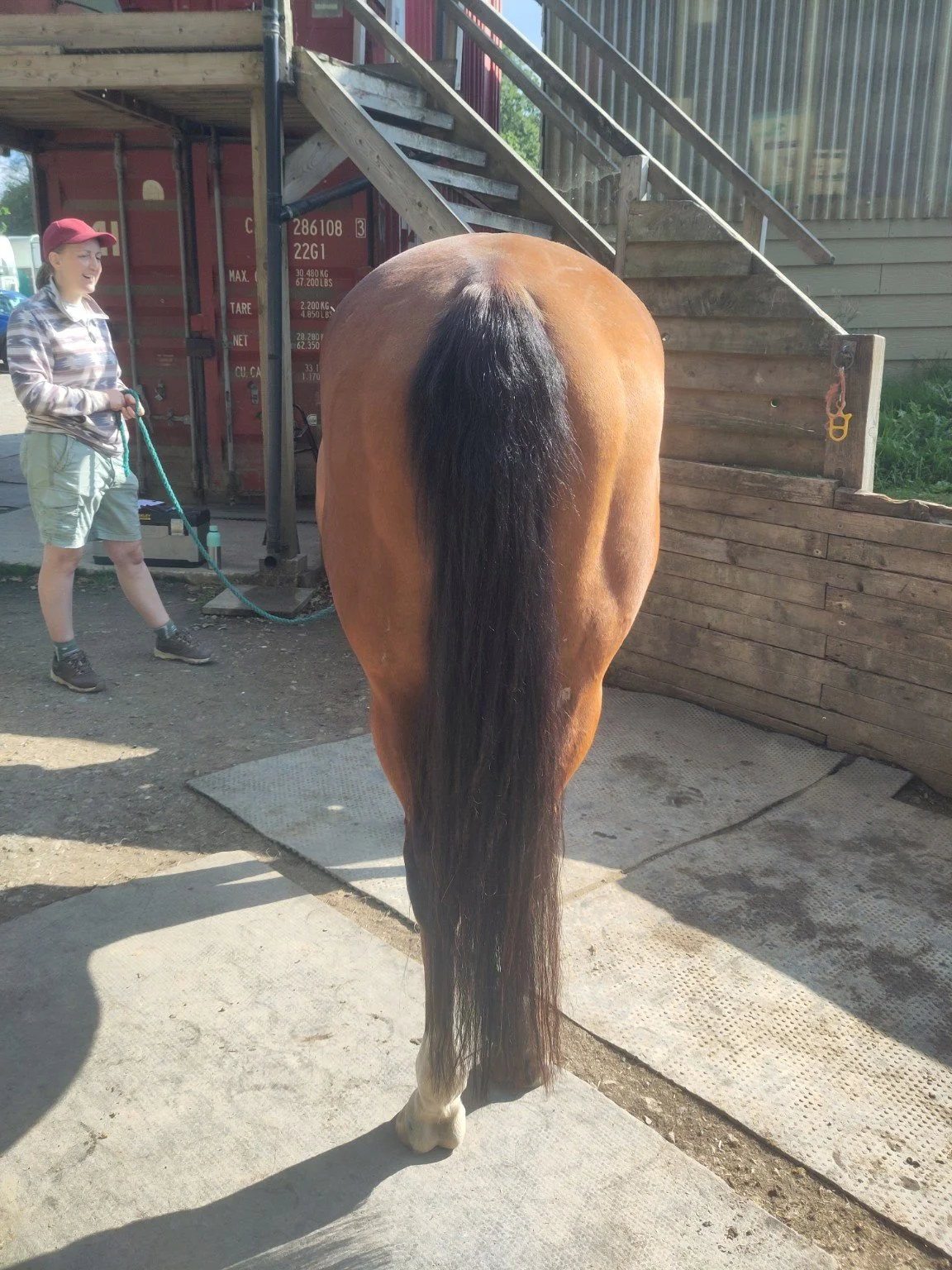
The photographs below were taken immediately before and after Cody's second treatment. They were not captured under controlled research conditions: the light, camera position and exact limb placement vary. They therefore cannot prove structural realignment.
What they do provide is useful supporting documentation. Across the different views, the post-treatment photographs show a more continuous outline through the neck, shoulder, thoracic wall, back and pelvis, with less visible global holding and superficial fascial tension.
What this case does and does not show
Cody's response is extremely encouraging, particularly because it occurred during his historically most symptomatic season. However, one case cannot establish that every headshaking horse will respond in the same way, or that osteopathic treatment alone caused the improvement.
Headshaking may involve trigeminal hypersensitivity, dental or sinus disease, ocular or ear discomfort, allergy and environmental irritation, tack or contact issues, gastric discomfort, cervical or cranial restriction, or several factors at once. Veterinary and dental investigation remain essential.
What Cody's case demonstrates is the value of looking beyond the symptom. Once significant pathology has been investigated appropriately, a detailed whole-horse assessment can identify mechanical and fascial relationships that may be contributing to the horse's overall sensitivity and ability to regulate.
Is your horse showing signs of headshaking?
If your horse is flicking the head, rubbing the nose, twitching, snorting excessively or reacting strongly to light, wind or exercise, the first step is careful documentation and appropriate veterinary assessment.
I work collaboratively with owners, veterinary surgeons, dentists and other professionals to assess complex cases and support the whole horse rather than offering a quick fix.
Contact Wild Voice to discuss your horse
This case study describes one horse's individual presentation and response. Osteopathic assessment does not replace veterinary diagnosis or treatment.